Difference between revisions of "Hamer-Focus"
(NMWIKI copy) |
(No difference)
|
Revision as of 19:57, 12 December 2009
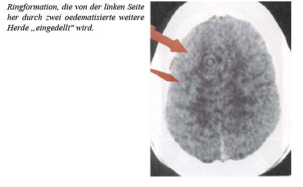
The former german physician Ryke Geerd Hamer and many followers of New Germanic Medicine count their diagnosis on a very particular interpretation of computer tomography brain scans of patients. This interpretation is not compatible with modern academic radiology. Hamer himself is not radiologist. In his books and on his webpages, he shows many brain scans but never shows details about the type of scanner used, exact date, or high voltage and exposure time used. He never shows the radiological findings and the reason why the scans were made. For a better understanding: In radiology, usually the left side of brain is shown on the right side, because a physician usually looks a patients into his face, standings before his feet. So he looks from downside to upside. Hamer presents his scans mirrored: the left side is seen on the left side of the picture.
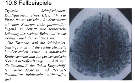
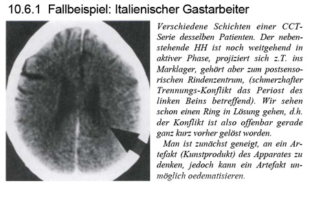
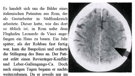
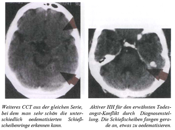
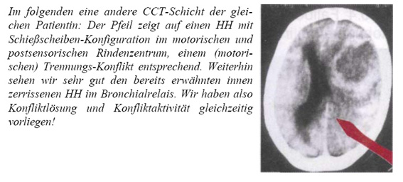
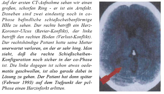
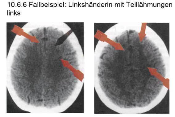
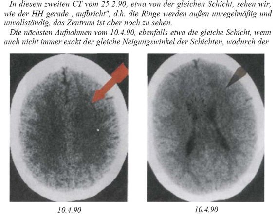
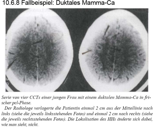
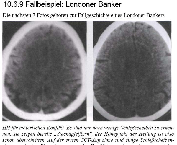
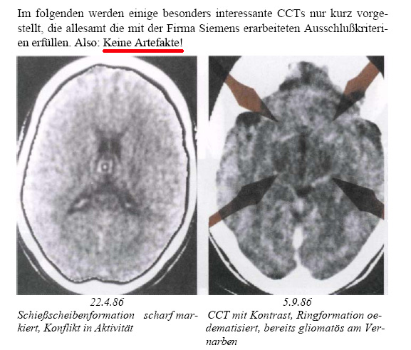
Hamer believes that a sudden shock-like onset of unforeseen so called biological conflict leads to a so-called "Dirk-Hamer-Syndrome" (DHS), which immediately (within a fraction of a second) produces a "cancer" in an organ. He postulates that every DHS-related process will take place synchronously in the brain, in the «organic brain» and in the organ. He adds that the forming tumours are allegedly controlled by the part of the brain that is ontogenetically connected with the organ in question. Hamer calls this the «ontogenetic system of tumours». In the brain, the biological conflict is said to give rise to the development of a so-called «Hamer focus». By this, Hamer means structures seen in CT brain scans which are shaped like a shooting target, or a single, mathematically precise circle. He claims that the locations of the Hamer foci and their degree of severity are correlated with the organs affected, the underlying biological conflict and the phase of the conflict. The non-radiologist Hammer adds also that radiology was not able to detect these Hamer-foci until his inventions in 1981. According to new medicine, the patient’s right- or left-handedness also plays a part.
The ring artefact (ring artifact)
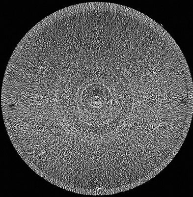
Artefacts were quite frequent at the beginning of computer tomography, and a particular type of artefact, the ring-artefact (or ring-artifact) [1] was sometimes seen in early CT scanners, especially those of third generation and are very seldom today with progess in manufacturing scanners. These artefacts are machinery-caused shapes superimposing the underlying scan. Usually they are easy to recognize and can be avoided by complying with the indications given by the manufacturer. Ring artefacts are always precise and perfect dark and bright concentric circles. Sometimes they may appear only as segments of a perfect ring and sometimes only one single circle can be seen (if a specific single sensor is defective or not calibrated). The center of the concentric circels corresponds to the axis of rotation of the scanner and may be seen sometimes outside of the subject or object in the scanner. These circles are caused or by a defectous single sensor, a sensor with different sensibility than the other sensors or after having forgotten to calibrate the whole scanner. An old fashion CT-scanner had to be calibrated (with air or a water-phantom) at the beginning of service. After some hours a message told the operator to stop scanning and to perform a new calibration procedure as temperature influences and other effects were modifying slowly the performance of the single sensors. These sensors must work at the limit of sensibility to avoid too large doses of radiation for the patient. Knowing this, an expert CT-operator may also be able to produce artificial rings-artefacts as he wants by avoiding calibration or by manipulating the device.
More stuff about this issue:
- http://radiographics.rsnajnls.org/cgi/content/full/24/6/1679
- http://www5.informatik.uni-erlangen.de/Lehre/WS0506/MB-JASS06/slides/h-1-6.pdf
The famous Siemens certificate december 22, 1989
Hamer and his followers often name a certificate of a german CT-manufaturer, the Siemens company dated december 22, 1989. And they believe that this paper would exclude the hypothesis that the Hamer-focus is in fact a tecnical artifact, namely a ring-artefact. But the opposite is true. This document can be used to identify ring-artefacts in many brain-scans shown by Hamer. This documents does not deal about new medicine or Hamer-focus.
It was Hamer himself to request this document (!) in 1989 that was underwritten the day december 22 [2]. Hamer was one of the two underwriting people, the other beeing a Siemens engineer (not a radiologist). Hamer was at that time already barred as a physician since three years and had to face the reproval by radiologists and other physicians to show artefacts. This document tells from the point of view of Siemens several conditions that are not compatible with ring artefacts.
Translation: Erlangen, 22.12.89. Possible ring artefacts. The underwriting [Hamer and engineer Feindor] have developped 8 [in fact only 7] excluding criteria excluding the presence of ring artefacts. Not a ring artefact is present if
- 1. in MRI (Magnetic resonance imaging or tomography) an anlogue structure is visible [at the same location]
- 2. the circles are not perfect circular, but show impressions, having a correlation with dislocation of tissue [irregular circles]
- 3. a formation corresponds to glial tissue [part of the brain, not beeing out of neurons. Glial tissue never shows up in circles however - see literature. Detection is only possible by analyzing a tissue sample]
- 4. the center or the circles do not correspond to the axis of rotation of the scanner ("parazentrale Schiessscheibenkonfiguration"="paracentral") [term used by Hamer]
- 5. other circles aside [not beeing therefore concentric] are seen, only one can be a ring artefact.
- 6. the circle-structures have a clinical course [history], in other words: if they will be seen in the same location in future CT scans, but looking different.
- 7. scanner dipendent artefacts are ring-shaped structures or ring segment shaped structures around the axis of rotation of the sanner. If these structures can be confused with biological structures, it is recommended to repeat the scan with a lateral or vertical dislocation of the patient. If the structure will not appear in a different location, in respect to known anatomical reference point, it is not an artefact.
ing Feindor, RG Hamer
This Siemens document does not mention new medicine or Hamer and does not exclude the fact that Hamer shows ring artefacts in his books. The precise circular shape of many structures showed by Hamer correspond to point 2 in this document. There are no neuropathological reports that any of the Hamer-focus has ever been identified to be of glial-tissue according to point 3.
expert opinion: prof. Maximilian Reiser (university of Munich) in 2007
To professor professor Maximilian Reiser (university of Munich) and president of german radiologists (Deutsche Röntgengesellschaft) were presented some CT brain-scans out of a book of Hamer. Reiser wrote in an expert opinion the day january, 22 2007:[3]
expert opinion of Prof. Dr. med. Dr. h,c, Maximilian Reiser, director of the institute of radiology for clinical radiology of the Ludwig-Maximilian-University Munich, president of the Deutsche Röntgengesellschaftät.
I willingly confirm you that the brain-scans presented in the "work" of Mr. Hamer have been interpreted in a completely inappropriate way by the author and are in clear conflict to scientifically justified knowledge and experience. An argumentative discussion with the content of Hamer's theories and the related interpretations of the brain scans is in my opinion not possible or lgoal-leading because Mr. Hamer is moving in a hermetically closed ambience of ideas and refuses any critic as expression of an arrogant "scolastic medicine". I would like to confim your corrections made in respect to some CT findings.
You may willingly cite this opinion as the opinion of the president of "Deutsche Röntgengesellschaft
With friendly regars, your M. Reiser
other opinions

The Swiss Study Group for Complementary and Alternative Methods in Cancer writes: ...The «Hamer foci» on the CT images in Hamer׳s books have been identified by radiological experts as typical artefacts produced by the radiological device which can appear in a poor-quality CT scan...[5] and the author Sören Ventegodt (Denmark) adds: ..The concentric circles in the Hamer focus, shown on the front page of his book[1], looks like an artefact and very little as a biological phenomena, which in humans are almost never seen as concentric circles... [6].
Warning letters to German radiologists
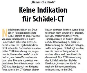
The Association of German Radiologists (Deutsche Röntgengesellschaft DRG) issued a warning letter to all members. The radiologists should know, that some people would try to undergo a CT-scan for further evaluation by GNM-therapists, violating German RÖV-regulation because lacking any stringend indication. The same warning was also issued by other German Medical Associations [7][8][9].
CT brain scans presented by R.G. Hamer
The pictures are from his book: [10].
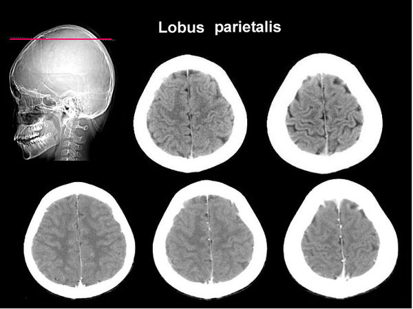
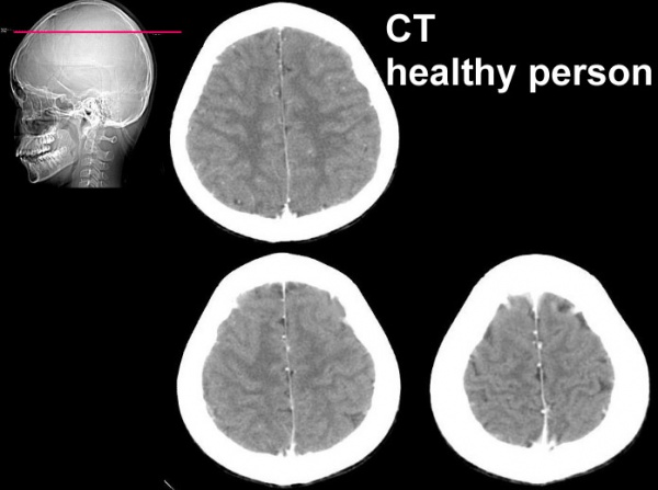
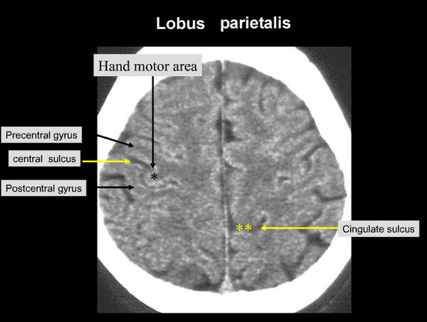
CT brain scans of a healthy subject to compare (not out of a Hamer-book)
A modern scanner is used here, picture-quality is far better.
 |
. |
 |
. |
 |
. |
Ring artefact example from a scientific study
other CT brains scans of Hamer-foci in the internet
references
- ↑ http://www.medcyclopaedia.com/library/topics/volume_i/r/ring_artefact/gring_artefact_fig1.aspx?s=ring%20artefact&scope=&mode=1
- ↑ *http://www.pilhar.com/Hamer/NeuMed/Zertif/891222.htm
- ↑ http://www.promed-ev.de/modules/news/article.php?storyid=105
- ↑ http://www.radpod.org/2008/03/24/ring-artefact-with-pseudomedical-interpretation/
- ↑ Swiss Study Group for Complementary and Alternative Methods in Cancer, SCAC. Hamer's «New Medicine» Document No. 01/02. [1]
- ↑ http://www.thescientificworldjournal.com/headeradmin/upload/2005.03.16.pdf
- ↑ http://www.kvsaarland.de/dante-cms/app_data/adam/repo/5975_CT_Untersuchungen_ohne_rechtfertigende_Indikation.pdf
- ↑ http://www.bezirksaerztekammer-trier.de/ak_aktuelles_det.php?lfd=280
- ↑ http://www.laekh.de/upload/Hess._Aerzteblatt/2009/2009_07/2009_07_05.pdf
- ↑ Hamer RG, Vermächtnis einer neuen Medizin, first part, editor. Amici Di Dirk


